Vesicle detection
Extracellular vesicles (EVs) are small particles surrounded by a lipid bilayer. Most cells are capable of secreting EVs. They carry cargos between cells and play roles in diverse processing from embryo development to injury response, and from angiogenesis to disease progression. The typical size of EVs is in the range from 50-1000 nm, and TEM is the most convenient instrument for their visualization. When analyzed by conventional TEM with heavy metal staining, most EVs exhibit a characteristic cup-shaped morphology. Here you can automatically detect and measure EVs in your images
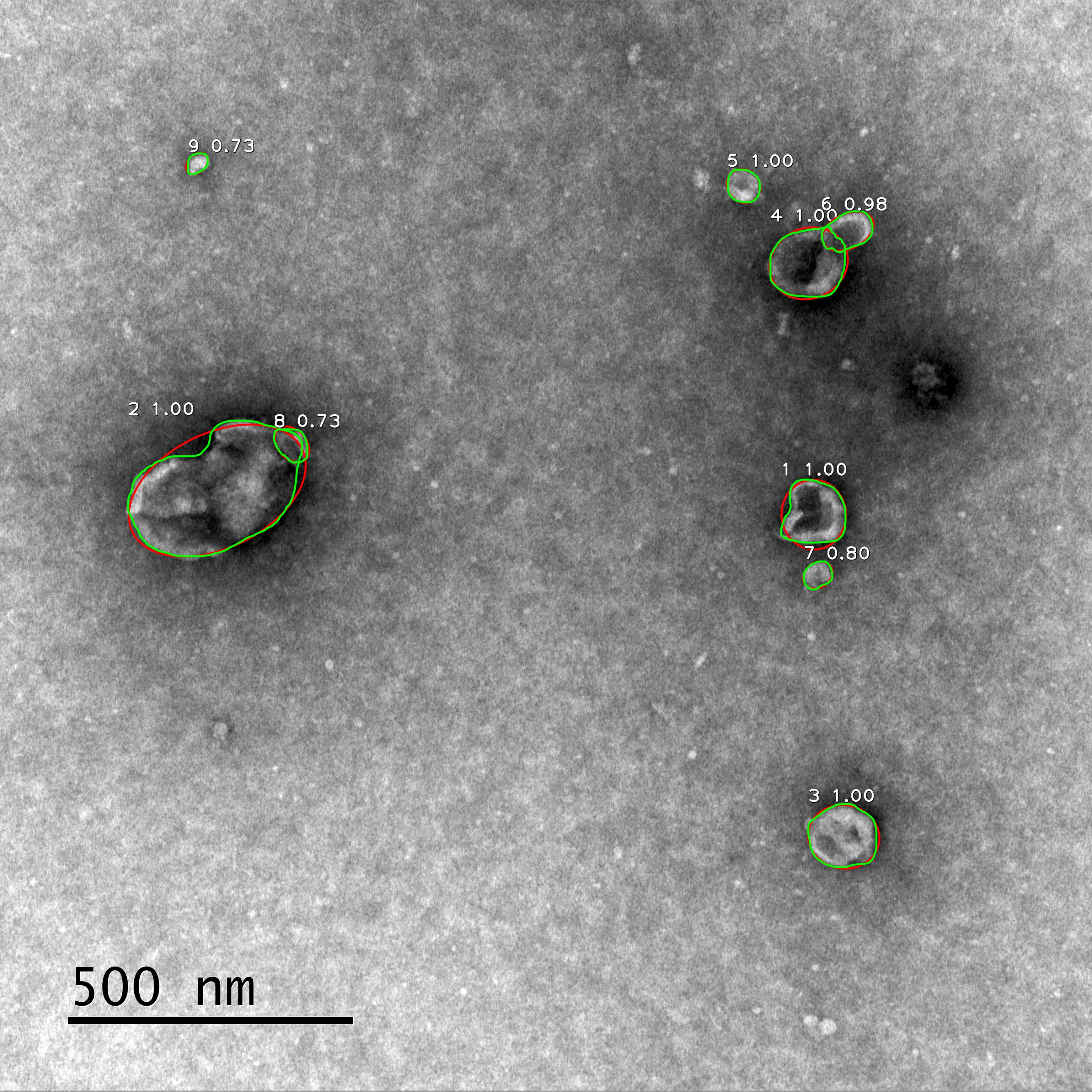
Detection is based on the Mask R-CNN. Each particle is identified, labeled, and measured. The green lines show the detected edges of the particles, and the red lines show the ellipses used for approximation.
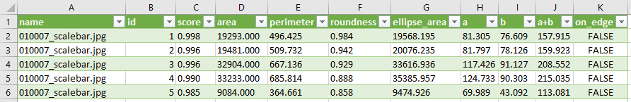
The measurement results are stored in a .csv file with the following parameters:
- Name – the file name
- Id – the particle number
- Score – the score of the particle, which is an estimate of the probability that this particle is a vesicle
- Area – particle area, measured in pixels
- Perimeter – particle perimeter, measured in pixels
- Roundness – a value in the range from 0 to 1, a measure of the particle roundness, calculated as Roundness = 4π(S/p2)
- Ellipse_area, a, b, and a+b are four parameters related to the ellipse approximating the detected particle. The values a and b are the semi-axes of the ellipse, and a+b is a relevant estimate of the particle size. All the parameters measured in pixels
- On_edge – a Boolean parameter indicating whether the particle touches the edge of the image
Dataset and code available here
Upload up to 10 images for detection
Supported 8 and 16 bit images in formats: png, jpg,
jpeg, tif, tiff
If you use ScanEV for your research, please cite the article "ScanEV – A neural network-based tool for the automated detection of extracellular vesicles in TEM images" by Igor Nikishin, Ruslan Dulimov, Gleb Skryabin, Sergey Galetsky, Elena Tchevkina, and Dmitry Bagrov // Micron, Vol. 145, 103044 (2021), doi: 10.1016/j.micron.2021.103044
This work was supported by RFBR, project №19-34-90148.